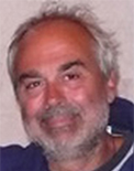
Curriculum vitae:
1976 Degree in Biological Sciences, University of Rome La Sapienza, 110/110 CL.
1977-1988 Professor of Sciences, Secondary State School.
1985 Department of Zoology, U. C., London, Research Fellowship (CNR-NATO)
1989-2010 Assistant Professor, Dept. of Biology, University of Rome “TorVergata”.
1992-1993 Dept. of Mol. Genetics, Ohio State University, Columbus, Visiting Professor (CNR fellowship)
1999 Dept. of Biology and Biochemistry, Center of Regenerative Medicine”, Bath University, UK , Visiting Professor (CNR fellowship)
2010-present Associate Professor, Dept. of Biology, University of Rome “Tor Vergata”.
Scientific activities
1975-1978Causal analysis of lens regeneration in X.laevis, University of Rome La Sapienza
1977-1983 Comparative Immunobiology in Urodela, Universityof Rome La Sapienza
1978-1980 Teaching Science, CNR-Comune di Roma for a new Zoo.
1985 Mitogenic antibodies to B cells surface immunoglobulin, Tumor Unit, Imperial Cancer Research Found, UC, London
1989-present Molecular and cellular aspects of regeneration (CNS, Eye, Limb)in Amphibians;
degenerative and regenerative processes in mammal muscle; tissue engineering and biomaterial technology.
Teaching activities
1978-1985 as Assistant Professor: General and Experimental Morphology, practical courses, University of Rome “la Sapienza”
1985-1986 as Term Professor: “Phylogenesis of immune system”, Sassari University
1989-1995 as Researcher: Comparative Anatomy, practical courses, University of Rome “Tor Vergata”
1995- 2010 as Assistant Professor: Experimental Morphology and Comparative Anatomy, University of Rome “Tor Vergata”
2010-present as Associate Professor: “Comparative Anatomy”,“Development and Regeneration”, “Regeneration and Stem Cells” courses,University of Rome “Tor Vergata”;
2000-present Member of PhD School, University of Rome “Tor Vergata”
2000 “Fondazione Roma” grant on the project: “Stemcellbasedapproaches to monogenic diseases”
Research topics:
The research projects of the lab will focus three main topics:
a) skeletal muscle tissue engineering; b) late stage muscular dystrophy recovery; c) amphibian and mammals limb regeneration.
a)Tissue engineering aims to mimic the environment where organogenesis took place and thus allow better stem cell engraftment and consequent new tissue histogenesis. Enhancing skeletal muscle regeneration and/or reconstruction using tissue engineering would be invaluable for the treatment of a variety of muscle diseases such as localized muscular dystrophies, post-traumatic or post-surgery tissue ablation and reconstruction of incontinent sphincters. Two general approaches are adopted to engineer skeletal muscle tissue: one is to isolate muscle progenitor cells from biopsies, expand and differentiate cells in a 3D defined environment in vitro (biomaterial or bioreactor) and reimplant the differentiated neotissue (in vitro tissue engineering). The second approach forecasts isolation of muscle stem cells, expansion in vitro and reimplantation of donor cells using a transport matrix, which promote myotube differentiation (myoblast transfer therapy). Moreover, muscle progenitor cells could be modified as vehicle for recombinant protein delivery (in vivo tissue engineering). Our efforts are focused on engineering skeletal muscle tissue by combining vessel associated myogenic progenitor cells (Mesoangioblast, Mabs) with the scaffold componentrepresented by PEG-Fibrinogen (PF), an injectable hydrogel. Our researchproject follows two main directions: in vivo myoblast transfer therapy for skeletal muscle tissue regeneration and in vitro tissue engineering for muscle reconstruction.
2D Mabs plastic culture shows myotube differentiation within 3-5 days after serum depletion, while using PF as scaffold for 3D Mabs culture,myofiber formation occurs in 24 hours, revealing the capacity of PF to promote and accelerate muscle differentiation in vitro. Moreover in vivo experiments conducted in a muscle injury mouse model (cardiotoxin crushed) using PF as carrier for Mabs injection demonstrated increased cell survival, ameliorated engraftment of transplanted cells likely due to improved integration and fusion of injected cells with host regenerating muscle fibers.
As for muscle reconstruction in vivo, we implanted PF and n-LacZ expressingMabs, modified to express the angiogenic factor Placental derived Growth Factor (Mabs-nLacZ/PlGF) subcutaneously intoimmunocompromised mice. We areattempting to implant PF loaded with Mabs-nLacZ/PlGF subcutaneously on the surface of the Tibialis Anterior of immunocompromised mice in order to investigate whether natural mouse TA movement could lead to parallel orientation and hence functionality of the artificial muscle; the next approach would be focus on tissue-engineer a muscle in a bigger mammal in order to reproduce a complete and functional piece of artificial muscle and grafting it to replace an injured, diseased or failing muscle.
b) Muscular dystrophies (MD’s) comprise a heterogeneous group of diseases characterized by primary muscle fibers wasting and progressive loss of strength, that in severe case likewise Duchene Muscular Dystrophy (DMD) leads to wheelchair dependence, respiratory failure and premature death (Emery 2002). In MD’s lacking or mutating on dystrophin or dystrophin-glycoprotein related members, protein complex linking muscle contraction machinery to the basal lamina, drives to muscle fragility and consequent myofibers degeneration accompanied by a severe inflammation with infiltration of immune cells. Degenerating fibers are repaired and replaced by myogenic resident stem cells (satellite cells), nevertheless dystrophic satellite cells produce muscle fibers prone to degeneration as well. Thus after several cycle of damage and repair, when they exhausted their regenerative capability, degenerating fibers are progressively substituted by connective and fatty tissue leading to a sclerotic environment with a remarkable microcirculation reduction and a severe chronic inflammation. This is the case of muscular dystrophy at advanced stages of the disease, representing the vast majority of affected individuals. Although molecular defect and ongoing mechanism of the disease are known, satisfactory therapy is not available, being still far from yielding complete skeletal muscle recovery.For pharmacological, gene and cell therapy, efficient delivery of the therapeutic drug, gene or cell to skeletal muscle represents the major challenge since skeletal muscle is the most abundant tissue in the body and is composed of multinucleated cells that do not divide. Furthermore, the pathological changes that occur in the advanced stages of the disease will reduce the efficacy of molecules, gene and cell delivery: mesoangioblasts (Mabs), vessel associated progenitor cells, treatment was greatly reduced when it was administered to mice older than two months of age. Even small molecules such oligonucleotides or drugs may loose efficacy when a dramatic reduction of the microcirculation may prevent their reaching the surviving muscle fibers. Therefore, the current trials that may be successful on young patients they would be unlikely to release a significant benefit when extended to cases at more advanced stages of the disease, the vast majority of affected individuals. We have recently demonstrated that tendon fibroblasts engineered to express a metalloproteinase and an angiogenic factor can restore microcirculation and reduce connective tissue deposition after intramuscular delivery to old dystrophic muscles. This treatment causes a dramatic amelioration of successive intra-arterial cell therapy that is restored to the same level of efficacy observed in young dystrophic muscle.
Hence in the light of above-mentioned results we are developing a new modular technology enabling a complete, groundbreaking regeneration of skeletal muscle. This technology consists of the combination of new generation of fibrinogenbiomaterial, the PEG-Fibrinogen, and mesoangioblasts. The field of regenerative medicine as a whole has faced two main obstacles: the first one is the difficulty, when using stem cells, of localizing the therapeutic effect to the target tissue in a specific manner. Previous clinical attempts of muscle regeneration in humans involving the resident myogenic precursors located underneath the muscle fiber basal lamina or satellite cells have failed partly because of their limited migratory capacity and survival. They tend to remain at the injection site vicinity, making it difficult to achieve an even distribution throughout the muscle and to go trough programmed cell death. Another reason for the failure of previous cell therapies is the fact that stem cells have not been protected adequately. The combination therapy we are attempting has not only proven to address these two obstacles when tested for its regeneration capacity in DMD animal models, but also showed superiority in the degree of regeneration obtained when compared to the mesoangioblasts alone.
c)Limb regeneration is one of the best examples of regeneration in vertebrates, since limb is a complex structure composed of different tissues. Urodele amphibians (newts and salamanders) are able to regenerate amputated limbs anytime during their life, while anuran amphibians (frogs) can regenerate their limbs during larval stages but progressively lose this capability during development until metamorphosis occurs. Mammals do not regenerate amputated limbs during adulthood but they possess the ability to regenerate digit tips during pre-natal and neonatal life. Limb regeneration in urodeles is due to the formation of an apical epithelial cap (AEC), which then induces the formation of a regenerative blastema. The blastema consists of a population of active proliferating progenitor cells and arises from dedifferentiation of stump tissues cells. It contains the intrinsic morphogenetic informations that are required to re-pattern the regenerating limb and direct the regenerative response. In spite of the deep knowledge on molecular mechanism occurring during amphibian limb regeneration processes the cellular movement, fate and commitment need to be elucidated yet. Since the idea is to focus on study Xenopuslaevis limb blastema cells identity. Recentlywe succeeded in elaborating a protocol for limb blastema stem cells isolation, growth and labelling. Limb blastema at different regeneration stages will be treated in order to obtain isolated stem cells; to understand their potentiality, freshly isolated blastema stem cells will be labelled (Lentiviral vector), implanted in regenerating and non regenerating amputated tadpole limb to analyse their capability in promote regeneration of all missing appendage or limited to specific tissue or structure related to their stage of origin and than to their differentiative state.
Mammals appendage regeneration is extremely reduced, during digit tip amputation in mice, a typical inflammatory response occurs. Fibroblasts begin synthesizing collagen and extracellular matrix proteins, which turn into a scar tissue. It hampers the intercellular signalling between the epidermis and the deeper tissues of the wound, avoiding the formation of an AEC and a blastema. That is one of the principle reasons why limb regeneration is impaired in adult mammals; in contrast, wounds in embryonic and foetal mammals heal without scar formation and involve a smaller inflammatory response, so that a partial regenerative response is possible. A “conservative” treatment in clinical cases of digit tip amputation in humans, by delaying the wound closure, in fact seems to promote a regenerative response.Since the idea to promote mouse digit tip regeneration pursuing three different approaches:
- cell therapy based on GFP labelled mesoangioblasts, vessel-associated progenitor cells, genetically modified for the expression of metalloproteinase-9 (Mabs-MMP9) in order to avoid scar tissue deposition and promote tissue regeneration;
- gene therapy based on third-generation lentiviral vectors to promote Dominant positive beta-Catenin (Dpbcat) expression which encodes for a dominant positive form of b-catenin in order to up regulate the Wnt/b-catenin pathway, it has been already demonstrated its necessary role during limb regeneration in amphibians by inducing blastema cell proliferation;
- biomaterial use (PEG-fibrinogen, PF) in order to allow a gradual and controlled viral vector release in vivo or to support the delivery of modified mesoangioblasts into the amputated digit tip.
The treatment of amputated digits of neonatalmice with Mabs-GFP/MMP9 results on formation of a loose cell layer whose morphology reminds an early blastema. This cellular cap could be due to the proliferation of the injected mesoangioblasts or it could appear after the mobilization of local progenitor cells or after the proliferation of dedifferentiated cells. Immunohistochemical analysis revealed that the injected mesoangioblast did not take part to the observed cellular cap in the treated mice.
In a similar way to the events of limb regeneration in amphibians, the up-regulation of Wnt/-catenin signalling pathway in the amputated digit tips of newborn mice leads to an increased proliferation, PF has been used in association with third-generation recombinant lentiviral vectors for the expression of Dpbcat in order to promote the same effect of Wnt/b-catenin pathway: the formation of a regenerative blastema or a blastema-like structure. The association of lentiviral vectors with biomaterials could increase their efficiency by protecting them from the surrounding environment and from the host immune response and also by slow releasing in loco.The use of biomaterials, like PEG-fibrinogen, in regenerative medicine could be promising because they are specifically designed to be degradable in vivo during the physiological processes of tissue remodelling and so they could behave like a scaffold for modified mesoangioblasts in cell-based therapies or for third-generation recombinant lentiviral vectors in gene therapy approaches.
Selected publications:
Bearzi C, Gargioli C, Baci D, Fortunato O, Shapira-Schweitzer K, Kossover O, Latronico MV, Seliktar D, Condorelli G, Rizzi R. lGF-MMP9-engineered iPS cells supported on a PEG-fibrinogen hydrogel scaffold possess an enhanced capacity to repair damaged myocardium. Cell Death Dis. 2014 Feb 13;5:e1053. doi: 10.1038/cddis.2014.12.
Montagna C, Di Giacomo G, Rizza S, Cardaci S, Ferraro E, Grumati P, De Zio D, Maiani E, Muscoli C, Lauro F, Ilari S, Bernardini S, Cannata S, Gargioli C, Ciriolo MR, Cecconi F, Bonaldo P, Filomeni G. S-nitrosoglutathionereductase (GSNOR) deficiency-induced S-nitrosylation results in neuromuscular dysfunction. Antioxid Redox Signal. 2014 Mar 31. [Epubahead of print]
Claudia Fuoco, Antonella Biondo, Emanuela Longa, Anna Mascaro, Keren Shapira-Schweitzer, M. Lavinia Salvatori, Sabrina Santoleri, Sergio Bernardini, Stefano M. Cannata, Roberto Bottinelli, DrorSeliktar, Giulio Cossu and Cesare Gargioli. In vivo generation of an entire, functionalskeletalmuscle. (EMBO Molecular Medicine under review)
Lettieri Barbato D, Aquilano K, Baldelli S, Cannata S, Bernardini S, Rotilio G, Ciriolo MR. (2014). Proline oxidase-adipose triglyceridelipasepathwayrestrains adipose celldeath and tissueinflammation.CELL DEATH AND DIFFERENTIATION, ISSN: 1476-5403, doi: 10.1038/cdd.2013.137. [Epubahead of print]
Dovere L, Fera S, Grasso M, Lamberti D, Gargioli C, Muciaccia B, Lustri AM, Stefanini M, Vicini E. The Niche-DerivedGlial Cell Line-DerivedNeurotrophicFactor (GDNF) Induces Migration of Mouse SpermatogonialStem/Progenitor Cells. PLoSOne. 2013 Apr 22;8(4)
Aquilano K, Baldelli S, Pagliei B, Cannata S, Rotilio G, Ciriolo M (2013). p53 orchestrates the PGC-1α-mediatedantioxidantresponseuponmild redox and metabolicimbalance. ANTIOXIDANTS & REDOX SIGNALING, vol. 18, p. 386-399, ISSN: 1523-0864, doi: 10.1089/ars.2012.4615
Fuoco C, Salvatori L, Biondo A, Shapira-Schweitzer K, Santoleri S, Antonini S, Bernardini S, Tedesco S, Cannata S, Seliktar D, Cossu G, Gargioli C (2012). Injectable PEG-fibrinogenhydrogeladjuvantimprovessurvival and differentiation of transplantedmyogenicprogenitors in acute and chronicskeletalmuscledegeneration .SKELETAL MUSCLE, vol. 2, p. 24-36, ISSN: 2044-5040, doi: doi: 10.1186/2044-5040-2-24.
Maiani E, Di Bartolomeo C, Klinger F, Cannata S, Bernardini S, Chateauvieux S, Mack F, Mattei M, De Felici M, Diederich M, Cesareni G, Gonfloni S (2012). Reply to: Cisplatin-inducedprimordialfollicleoocytekilling and loss of fertility are notprevented by imatinib.. NATURE MEDICINE, vol. doi:, p. 1172-1174, ISSN: 1078-8956, doi: 10.1038/nm.2852.
Garcia PS, Chieppa G, Desideri A, Cannata S, Romano E, Luly P, Rufini S. (2012). Sticholysin II: A pore-formingtoxinas a probe torecognizesphingomyelin in artificial and cellularmembranes.. TOXICON, vol. Epubahead of print, ISSN: 0041-0101
Rizzi R, Bearzi C, Mauretti A, Bernardini S, Cannata S, Gargioli (2012). Tissueengineeringfor skeletalmuscleregeneration. MUSCLES, LIGAMENTS AND TENDONS JOURNAL, vol. 2, p. 230-234, ISSN: 2240- 4554.
Fuoco C, Cannata S, Bottinelli R, Seliktar D, Cossu G, Gargioli C (2012). autologous progenitor cellsin ahydrogelform a supernumerary and functionalskeletalmuscle in vivo. JOURNAL OF TISSUE ENGINEERING AND REGENERATIVE MEDICINE, vol. 6, p. 116, ISSN: 1932-7005m.
Fuoco C, Cannata S, Bottinelli R, Seliktar D, Cossu G and Gargioli C. Autologous progenitor cellsin ahydrogelform a supernumerary and functionalskeletalmuscle in vivo. Journal Of TissueEngineering And Regenerative Medicine. Sep 20126; (1): 116
Rizzi R, Bearzi C, Arianna M, Bernardini S, Cannata S and Gargioli C.Tissue engineering for skeletal muscle regeneration. MLTJ 2012; 2(3): 230-34
Fuoco C, Biondo A, Salvatori ML, Shapira-Schweitzer K, Santoleri S, Bernardini S, Cannata S, Seliktar D, Cossu G and Gargioli C. InjectablePEG-fibrinogenimprovessurvival and differentiation of transplantedmyogenicprogenitors in acute and chronicskeletalmuscledegeneration. SkeletMuscle 2012; 26;2(1):24. [Epubahead of print]
Pichavant C, Gargioli C, TremblayJP.IntramuscularTransplantation of Muscle Precursor Cells over-expressing MMP-9 improvesTransplantation Success. PLoSCurr. 2011 Oct 26;3:RRN1275.
Hamza L, Girardi T, Castelli S, Gargioli C, Cannata S, Patamia M, Luly P, Laraba-Djebari F, Petruzzelli R, Rufini S. Isolation and characterization of a myotoxin from the venom of Macroviperalebetinatransmediterranea.Toxicon 2010 Apr 14 [Epubahead of print]
Bernardini S, Gargioli C, Cannata SM and Filoni S. Neurogenesisduringoptictectumregeneration in Xenopuslaevis.DevGrowth 2010 May;52(4):365-76
Bernardini S, Gargioli C, Cannata SM and Filoni S. The effect of retinyl-palmitateon brain regeneration of larvalXenopusLaevis.Italian Journal of Zoology 2009, ISSN: 1125-00033-8
Gonfloni S, Di Tella L, Caldarola S, Cannata SM,Klinger Fg, Di Bartolomeo C, Mattei M, Candi E, De Felici M, Melino G, Cesareni G. (2009). Inhibition of the c-Abl-TAp63 pathwayprotects mouse oocytes from chemotherapy-induceddeath. NATURE MEDICINE, vol.15, p.1179-1185, ISSN:1078-8956.
Gargioli C, Giambra V, Santoni S, Bernardini S, Frezza D, Filoni S and Cannata S M. The lens-regeneratingcompetence in the cornea and epidermis of larvalXenopuslaevisisrelated to pax6 expression. J Anat. 2008 May 212 (5): 612-20.
Scardigli R, Gargioli C, Tosoni D, Borello U, Sampaolesi M, Sciorati C, Cannata SM, Clementi E, Brunelli S and Cossu G. Binding of sFRP-3 to EGF in the extra-cellularspaceaffectsproliferation, differentiation and morphogeneticeventsregulated by the twomolecules. PLoS ONE. 2008 Jun 18;3(6):e2471.
Gargioli C, Coletta M, De Grandis F, Cannata SM, and Cossu G. PlGF-MMP9 expressingcellsrestoremicrocirculation and efficacy of celltherapy in olddystrophicmuscle. NatMed. 2008 Sep 14(9):97
Research group and contact:
Stefano M. Cannata:ProfessoreAssociato, Tel. +39 06 72594815, Fax. +39 06 2023500, Email cannata@uniroma2.it
Cesare Gargioli:Ricercatore TD, Tel. +39 06 72594815, Fax. +39 06 2023500 Email cesare.gargioli@uniroma2.it
Sergio Bernardini:Funzionario Tecnico, Tel. +39 06 72594816, Fax. +39 06 2023500, Email Sergio.bernardini@uniroma2.it
Stefano Testa: Dottorando, Tel. +39 06 72594816, Fax. +39 06 2023500, Email stefanotesta87@hotmail.it


Università di Tor Vergata